Shoulder Tendons Chart - Labeled Anatomy Chart Of Shoulder Ligaments On White Background Stock Photo Download Image Now Istock - Muscle general anatomy 12 photos of the muscle general anatomy general anatomy of a muscle, general anatomy of muscle fibers, general anatomy of skeletal muscle, muscle general anatomy, muscle general anatomy ppt, human muscles, general anatomy of a muscle, general anatomy of muscle fibers, general anatomy of.. The etiology is most of the time traumatic and related either to sport or accidents. At the shoulder joint, there are actually two tendons that connect the biceps to the bone, which is why the muscle is called the biceps: The subacromial bursa reduces friction beneath the deltoid, promoting free motion of the rotator cuff tendons. It appears in a convenient poster size 50 x 67 cm (20x26'') and can be written on and wiped off with non permanent markers. Each illustration is clealy labeled and injuries are textually described.
This page is about shoulder tendons chart,contains what is causing your shoulder pain? This anatomical chart presents useful information about the shoulder and elbow. Typically, a shoulder may need. It stabilizes the shoulder and holds the head of the humerus in the. Please understand that our phone lines must be clear.
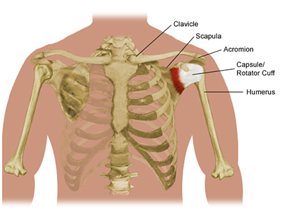
Overview Of Shoulder Problems And Treatments Piedmont Healthcare from www.piedmont.org Bi, meaning two, attachments at the shoulder. Each illustration is clealy labeled and injuries are textually described. The condition causes pain and tenderness just outside a joint. The main function of the rotator cuff is to keep the ball of the upper arm bone within the shoulder socket. Anterior view showing muscles, bones, liagments, nerves, veins and arterires Shoulder tendinopathy is an injury to the shoulder tendons. Related posts of shoulder muscles and tendons diagram back muscle diagram & pain. The rotator cuff muscles and tendons may be injured by trauma, such as falling when skiing or biking, or from arthritic spurs that form within the shoulder and erode the cuff tissue over time.
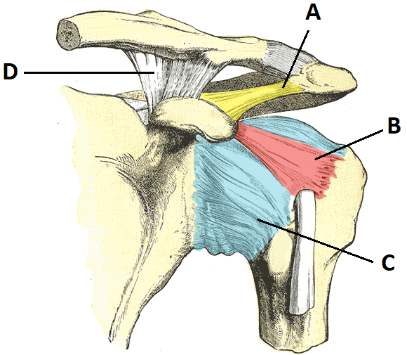
The cuff part comes from the four tendons merging together to form a cap or hood around the head of the.
It reduces wear and tear on the tendon during movement at the shoulder joint. Or a broken collarbone most often happens from falling and landing directly onto your shoulder, such as while bicycling, skiing, or. Your rotator cuff consists of the muscles and tendons in your shoulder. The collection of muscles and tendons in the shoulder is known as the rotator cuff. Anterior view showing muscles, bones, liagments, nerves, veins and arterires The etiology is most of the time traumatic and related either to sport or accidents. How tendons and connective tissue work. Dislocated shoulder, broken bone (such as the upper arm or collarbone), torn or ruptured tendon pain on top of the shoulder (where the collarbone and shoulder joint meet) problems in the acromioclavicular joint, like dislocation or stretched or torn ligaments It appears in a convenient poster size 50 x 67 cm (20x26'') and can be written on and wiped off with non permanent markers. Learn vocabulary, terms and more with flashcards shoulder joint is formed by a group of ligaments that connect humerus to glenoid. The cuff part comes from the four tendons merging together to form a cap or hood around the head of the. Muscle general anatomy 12 photos of the muscle general anatomy general anatomy of a muscle, general anatomy of muscle fibers, general anatomy of skeletal muscle, muscle general anatomy, muscle general anatomy ppt, human muscles, general anatomy of a muscle, general anatomy of muscle fibers, general anatomy of. The muscles are attached to the bones by tendons.
This anatomical chart presents useful information about the shoulder and elbow. In either case, the repeated motion can lead to rotator. It stabilizes the shoulder and holds the head of the humerus in the. Other causes are degenerative joint disease and arthritis. How tendons and connective tissue work.
Injuries Of Shoulder Exam Room Anatomy Posters Clinicalposters from cdn.shopify.com Inflammation of the bursa) can be a cause of shoulder pain. The subacromial bursa lies between the rotator cuff and shoulder blade and protects the tendons in this area. Muscle charts female muscle mini. (less common) tendinosis — tiny tears in the tendon with no significant inflammation. The most commonly affected tendons in the shoulder are the four rotator cuff tendons and one of the biceps tendons. In each case it is important to be familiar with some basic examination tools that can help us confirm the presence of a shoulder lesion. A tendon is a structure that connects muscle to bone, and the biceps are connected by tendons at both the elbow and shoulder joints. Related posts of shoulder muscles and tendons diagram back muscle diagram & pain.
Shoulder tendonitis is inflammation of your rotator cuff or bicep tendons, often caused by overuse of the arms such as in baseball, weight lifting, and racket sports.
If you tear your biceps tendon at the shoulder, you may lose some strength in your arm and have. Shoulder tendons chart ~ labeled anatomy chart of shoulder ligaments on white background stocktrek images. For instance, professional baseball players, swimmers, tennis players, and golfers are susceptible to tendinitis in their shoulders, arms, and elbows. The rotator cuff is a group of muscles and tendons that surround the shoulder joint, keeping the head of your upper arm bone firmly within the shallow socket of the shoulder. Calcific tendonitis occurs when calcium crystals are deposited within a tendon, most commonly within the rotator cuff tendons. It stabilizes the shoulder and holds the head of the humerus in the. These are located in the shoulder blade area, and each related tendon also attaches to the humerus. Tendons are thick fibrous cords that attach muscles to bone. The pain is likely caused by impingement of the tendons or bursa in that area of your shoulder. This inflammation to the tendons in shoulder impingement syndrome is a condition where rotator cuff tendons of the shoulders are. The cuff part comes from the four tendons merging together to form a cap or hood around the head of the. The most commonly affected tendons in the shoulder are the four rotator cuff tendons and one of the biceps tendons. Back muscle diagram & pain 12 photos of the back muscle diagram & pain back muscle diagram, back muscle diagram exercise, back muscle diagram pain, back muscles diagram a comprehensive view, back muscles diagram and ligaments, human muscles, back muscle diagram, back muscle diagram exercise, back muscle.
Your rotator cuff helps provide shoulder motion and stability. Or a broken collarbone most often happens from falling and landing directly onto your shoulder, such as while bicycling, skiing, or. The subacromial bursa lies between the rotator cuff and shoulder blade and protects the tendons in this area. Each illustration is clealy labeled and injuries are textually described. The upper arm bone (the humerus) is connected to the shoulder by muscles and tendons.
The Shoulder Joint Structure Movement Teachmeanatomy from teachmeanatomy.info Please understand that our phone lines must be clear. The muscles are attached to the bones by tendons. They connect your upper arm bone to your shoulder blade. Rotator cuff and shoulder conditioning program introduction 1 additional notes purpose of program _____ after an injury or surgery, an exercise conditioning program will help you return to daily activities and enjoy a more active, healthy lifestyle. Anatomy and injuries of the shoulder illustrates the following normal anatomy: Inflammation of the bursa) can be a cause of shoulder pain. The subacromial bursa lies between the rotator cuff and shoulder blade and protects the tendons in this area. The cuff part comes from the four tendons merging together to form a cap or hood around the head of the.
A detailed chart showing normal anatomy of the shoulder as well as common injuries.
Shoulder tendinitis occurs as a result of sports injuries, by repetitive use or overuse of the tendons, or from a sudden, more serious injury. The supraspinatus, the infraspinatus, the teres minor and the subscapularis. It appears in a convenient poster size 50 x 67 cm (20x26'') and can be written on and wiped off with non permanent markers. As people age and are less active, tendons start to degenerate and lose strength. Learn vocabulary, terms and more with flashcards shoulder joint is formed by a group of ligaments that connect humerus to glenoid. The cuff part comes from the four tendons merging together to form a cap or hood around the head of the. Shoulder pain is one of the most common complaints in the outpatient setting. Rotator cuff and shoulder conditioning program introduction 1 additional notes purpose of program _____ after an injury or surgery, an exercise conditioning program will help you return to daily activities and enjoy a more active, healthy lifestyle. This inflammation to the tendons in shoulder impingement syndrome is a condition where rotator cuff tendons of the shoulders are. The main function of the rotator cuff is to keep the ball of the upper arm bone within the shoulder socket. A tendon is a structure that connects muscle to bone, and the biceps are connected by tendons at both the elbow and shoulder joints. This page is about shoulder tendons chart,contains what is causing your shoulder pain? The condition causes pain and tenderness just outside a joint.
Shoulder Tendons Chart - Labeled Anatomy Chart Of Shoulder Ligaments On White Background Stock Photo Download Image Now Istock - Muscle general anatomy 12 photos of the muscle general anatomy general anatomy of a muscle, general anatomy of muscle fibers, general anatomy of skeletal muscle, muscle general anatomy, muscle general anatomy ppt, human muscles, general anatomy of a muscle, general anatomy of muscle fibers, general anatomy of.. There are any Shoulder Tendons Chart - Labeled Anatomy Chart Of Shoulder Ligaments On White Background Stock Photo Download Image Now Istock - Muscle general anatomy 12 photos of the muscle general anatomy general anatomy of a muscle, general anatomy of muscle fibers, general anatomy of skeletal muscle, muscle general anatomy, muscle general anatomy ppt, human muscles, general anatomy of a muscle, general anatomy of muscle fibers, general anatomy of. in here.